California Startup, Tribogenics, Develops Smart Phone Sized Portable X-ray Machines
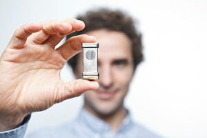
A Southern California startup called Tribogenics is using a new method of producing X-rays, fleshed out by Darpa-funded research at UCLA, to make X-ray machines more robust and portable.

Share
Doctors and dentists take millions of X-rays globally each year. Governments use X-rays to scan luggage and shipping containers for hazardous materials. Yet the technology underlying X-ray machines hasn’t changed much in the last hundred years.
We still accelerate electrons through glass vacuum tubes and slam them into material at the end to produce X-rays. The machines are bulky, fragile, and expensive.
But that may not hold true much longer. A California startup called Tribogenics is using a novel method of producing X-rays, fleshed out by Darpa-funded research at UCLA, to make X-ray emitters sturdier and more portable.
The firm derives its name from a natural process called triboluminescence. When some materials—tape, sugar, or famously, Wint-O-Green Life Savers—are pulled apart, positive and negative charges separate and, like lightning, reunite in a flash of light.
Triboluminscence has long been observed by scientists, but in 2008, it got a practical use. In a paper published in Nature, UCLA researchers produced X-rays in addition to visible light by simply unrolling Scotch tape in a vacuum.
Using no more than a piece of dental film, two rolls of tape, and a vacuum chamber, the scientists produced an X-ray image of a finger.
One of those researchers, Carlos Camara, now chief scientist at Tribogenics, noted the machines dentists use are orders of magnitude larger and more complex. “This is the cheapest way to generate X-rays at this scale,” Camara said.
Now, with almost $9 million in funding from the likes of Peter Thiel’s Founders Fund, Tribogenics is aiming to use triboluminescence to shake up X-ray tech.
Exploiting the same principle behind those early Scotch tape experiments, the firm says they’ve developed a new “metal-polymer” technology that can be embedded in comparatively tiny devices. Users will be able to employ the devices by themselves (as a point source) or together in an array.
Further, according to Tribogenics, “By eliminating the need for high voltage and fragile glass tubes, our solutions are safe to use in any environment.” And of course, all this also makes them much more portable.
Earlier this year, Tribogenics collaborated with Los Alamos National Laboratory to build a prototype device using their X-ray source. The MiniMAX (Miniature, Mobile, Agile, X-ray) camera weighs in at less than five pounds and was used to image the insides of a calculator.
Be Part of the Future
Sign up to receive top stories about groundbreaking technologies and visionary thinkers from SingularityHub.


Scott Watson of Los Alamos said, “We designed MiniMAX to demonstrate that such a system will open up new applications in security inspection, field medicine, specimen radiography and industrial inspection."
Meanwhile, Tribogenics is working on their own portable X-ray device, called the Pocket XRF. The device is designed for recycling and precious metal identification, but the firm is also developing devices for medical diagnostics and therapy.
The UCLA team's Juan Escobar saw the potential early on when he said, “You could have these devices in third world countries where electricity is a very expensive commodity. Where it’s not that easy to get an X-ray.” Indeed.
Ever since James T. Kirk first scanned M-Class planets for sexy aliens, we've had this dream of a multipurpose, handheld scanner like Kirk's tricorder—a magical, portable device to analyze the structure and state of inanimate objects and biological beings.
The Qualcomm Tricorder X Prize, whose official field of 34 competitors was recently announced, hopes to deliver a tricorder-like device into human hands in the coming years. (Check out our previous coverage of Tricorder X here.)
In the future, a mobile device (perhaps simply a smartphone) might draw data from a small array of portable external sensors.
These might include lab-on-a-chip devices to analyze small amounts of blood, urine, and saliva; sensors to test vital signs like blood pressure, heart rate, and respiration; and perhaps, as Tribogenics hones their design, a small X-ray emitter.
Diagnostically, this would be a key addition. Most commonly, X-rays image fractures or thinning of bones (osteoporosis) and cavities. They can also help diagnose lung and heart diseases, tumors, and gall and kidney stones.
Most in developed countries can visit a clinic with an X-ray machine. That's rarely the case in remote developing areas. The power of mobile medicine to serve the underserved may, in coming years, be greater (and less magical) than we imagine today.
Image Credit: Banner image courtesy of Shutterstock.com; body image courtesy of Tribogenics
Jason is editorial director at SingularityHub. He researched and wrote about finance and economics before moving on to science and technology. He's curious about pretty much everything, but especially loves learning about and sharing big ideas and advances in artificial intelligence, computing, robotics, biotech, neuroscience, and space.
Related Articles

More Space Junk Is Plummeting to Earth. Earthquake Sensors Can Track It by the Sonic Booms.

This ‘Machine Eye’ Could Give Robots Superhuman Reflexes

Elon Musk Says SpaceX Is Pivoting From Mars to the Moon
What we’re reading


