Your Doctor Can Now Examine an Exact 3D Replica of Your Heart in Virtual Reality

Share
Not so long ago, the only way to see what was going on inside a person’s body involved something sharp. Now, we can image the body’s organs without a single incision.
However, though we’ve made huge strides in medical imaging, it’s still far from perfect. One of the biggest challenges? Patients are three-dimensional, but medical images are flat; they each show a different slice of the anatomy in question. This makes gaining a volumetric understanding of what you’re looking at—which is critical when dealing with anatomy—a key skill.
To get a good picture of what they’re dealing with, doctors have to imagine the actual 3D organ by mentally piecing together all these flat images. This isn’t easy, and some doctors are better at it than others. In short, they’re preoccupied with a visualization problem—a process that is prone to errors, wastes time and causes fatigue for physicians—instead of concentrating on solving a clinical problem—their primary and most important job.
Take surgical planning, for example. Today, radiologists look through hundreds and hundreds of flat images, and then draw a diagram (yes, by hand) to show the surgeon how to approach a given procedure. Then the surgeon operates on the patient with no advance knowledge of his or her actual volumetric anatomy.
One surgeon I spoke with summed it up perfectly: “I’ve never opened up a patient and seen a 2D view!” Another surgeon specializing in image-guided surgery told us that “half the time I am guessing” when navigating 3D anatomy using 2D images.
Sounds a little scary, no? Never fear. It won’t be like this much longer.
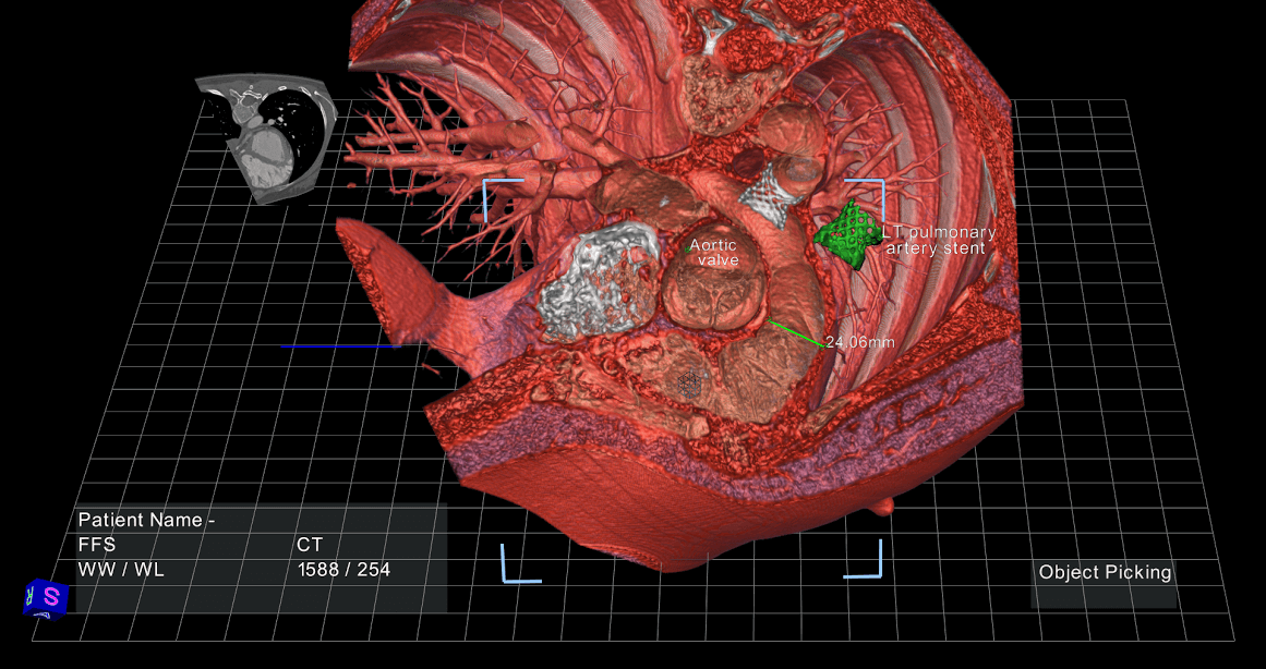
There’s a pretty powerful solution just now arriving—advanced image rendering through interactive virtual reality. EchoPixel (my company) uses virtual reality to help doctors visualize each patient’s unique anatomy and internal structure in a floating 3D image. The software uses DICOM data, which is already embedded in every MRI scan, CT scan, or ultrasound image.
Here’s how it works:
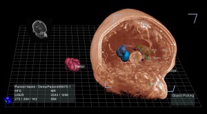
You stand at a workstation wearing a pair of 3D glasses. (Our software works with any VR hardware device, but we’re currently partnered with Zspace for the display.) A 3D image of an organ floats in front of you, an exact replica of a patient’s heart, colon, or brain. Using a stylus, you can turn, dissect, zoom, remove pieces—basically manipulate the image however you want.
It’s about as close as you can get to visualizing and interacting with the real thing.
Indeed, medical imaging is an ideal early use-case for virtual reality, because these volumetric imaging datasets already exist (unlike in entertainment, for example, where developers will have to create new content to run on VR devices). If we can help solve the 3D problem, then doctors can refocus their energy and expertise on diagnosis and treatment.
We see a huge opportunity for this sort of imaging technology to shape the way doctors work.
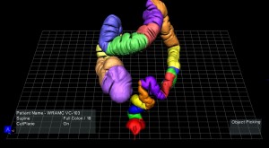
For example, the virtual colonoscopy procedure is becoming a popular alternative to the dreaded optical colonoscopy (recommended as a regular procedure for every person over age 50). Instead of requiring total sedation and a full day of recovery, the virtual procedure allows the doctor to examine a CT scan of the colon to identify any potentially cancerous lesions.
Even President Obama recently opted for this less invasive virtual procedure.
Even so, it’s not easy on doctors. They have to scan through hundreds of flat images, trying to identify cancerous lesions. It generally takes 30-40 minutes, is mentally fatiguing, and carries significant risk of missing a dangerous growth.
Using virtual reality, on the other hand, a doctor can “fly through” each segment of the colon in 3D space. The difference is extraordinary. One of the nation’s top specialists at UCSF (who we’re working with currently) is completing the procedure in just 5-10 minutes using interactive VR.
And she’s finding her ability to correctly identify cancerous lesions is up a full 100%.
Be Part of the Future
Sign up to receive top stories about groundbreaking technologies and visionary thinkers from SingularityHub.

In another example, we’ve been working with specialists at a leading implantable device manufacturer on a procedure to implant a heart device for atrial fibrillation. This device helps prevent blood clots from entering the bloodstream in certain patients at high risk for strokes.
The challenge here is determining the correct size for the device. Generally, it requires several iterations to implant the correctly sized device. Sizing can take up to 45 minutes, and still results in a 2.7 device per implant ratio with a cost of $20,000 for each incorrectly sized implant.
However, using an accurate 3D image takes away much ambiguity in sizing the device. As a result, doctors were consistently completing the procedure in just two minutes! Once again, virtual reality is proving to be not just an aid, but a complete game-changer in the operation.
We’re at the early stages, but are seeing similar results across a range of procedures that rely on medical images. With the ability to more fully understand a patient’s anatomy, doctors can produce better outcomes for patients in less time, which not only benefits the doctors themselves, but significantly reduces risks to the patient in question.
And this is just the beginning.
Imagine that doctors and medical students could practice surgery multiple times in VR before ever stepping into an operating room, following a step-by-step procedure from the best specialist in the world. Or a patient could carry around a digital file of their anatomy, with localized annotations from different specialists they had visited.
Instead of talking to each other with hand-drawn diagrams, now radiologists and surgeons can plan an operation together using a 3D image, making digital notes on key structures to operate on (and just as crucially, to avoid). This kind of communication “gap” is common among healthcare professionals, and we see VR technology as a communications tool as well.
Most people now agree that digital medicine is the future of health. But that doesn’t just mean numbers in a spreadsheet. We think virtual reality has a huge role to play in the healthcare industry, and it’s just getting started.
Ron Schilling, PhD, is the CEO of EchoPixel. Dr. Schilling has 35 years of operating and general management experience in the medical device and technology industries, at Toshiba, Diasonics and General Electric. He also teaches business strategy at Stanford and serves on several corporate boards in the medical field. You can learn more about Echopixel here.
To get updates on Future of Virtual Reality posts, sign up here.
Image credit: Shutterstock.com and Echopixel
Dr. Schilling has 35 years of operating and general management experience in the medical device and technology industries, at Toshiba, Diasonics and General Electric. He was a director at Stentor at 2007 when it was sold to Philips. He also teaches business strategy at Stanford and serves on several corporate boards in the medical field. Dr. Schilling earned his B.SC. EE from City College of New York, his M.Sc. from Princeton University, and a Ph.D. from the Polytechnic Institute of New York University.
Related Articles

Space Launch Costs Have Fallen 96% Since 1960. They Aren’t Done Yet.
")
This Week’s Awesome Tech Stories From Around the Web (Through July 18)

Anthropic Says Chatbots Have What May Be a Key Feature of Consciousness. Are They Right?
Space Launch Costs Have Fallen 96% Since 1960. They Aren’t Done Yet.
This Week’s Awesome Tech Stories From Around the Web (Through July 18)
Anthropic Says Chatbots Have What May Be a Key Feature of Consciousness. Are They Right?
What we’re reading