This MIT Device Maps the Human Brain With Unprecedented Resolution and Speed
Share
A squishy, fatty, beige-colored organ covered with grooves and ridges, the brain doesn’t look all that impressive on the surface.
But hidden underneath are up to 100 billion neurons and 100 trillion synapses—the connections between neurons that form networks—densely packed in a squishy three-pound organ that controls our thoughts, feelings, movement, memories, and sense of self.
For the past two decades, scientists have carefully dissected the internal neural connections and workings of the brain by carefully chopping it up into paper-thin pieces. From there, they’ve built multiple maps of the brain’s cellular population, architecture, connections, and gene expression. Like charting the landscape of a new world, these maps have been consolidated into what amounts to a Google Maps for the brain. These atlases allow us to decipher brain function, bridging genetic expression to cell functions, network connections, and behavior.
At least for rodents and other animals. Mapping the brain is incredibly difficult and time-consuming. A small chunk of a mouse’s brain, when imaged at single-cell resolution, takes years to process, scan, and reconstruct into 3D computer models. Any trip-ups during the process ruins the product. Mapping the human brain, much larger in size, is far more difficult.
This month, a team from MIT developed a “holistic” brain-mapping platform that captures the anatomy of large slices of the human brain with unprecedented resolution and speed, slashing a process that normally takes between a week and a month to a few days.
They used the platform to image an Alzheimer’s brain, after physically expanding brain tissues with a hydrogel. The automated system sliced, imaged, and automatically stitched the images together and found myriads of cellular changes and problems with neural connections, including inflammation.
Compared to previous brain mapping projects, which often require months or years, the new platform mapped different levels of the brain’s physical makeup—from synapses to local neural circuits and brain-wide connections in slabs of human brain tissue—in just a few days.
“We performed holistic imaging of human brain tissues at multiple resolutions from single synapses to whole brain hemispheres, and we have made that data available,” study author Kwanghun Chung said in a press release.
To be clear, the technology has only been used on slabs of human brain tissue and hasn’t yet charted the entire brain’s neurons and connections. But “this technology pipeline really enables us to analyze the human brain at multiple scales. Potentially this pipeline can be used for fully mapping human brains,” said Chung.
Three-Way Upgrade
Here’s how brain mapping technology usually works. Whole brains are sliced into wafer-thin pieces on a machine called a vibratome—think of it as a souped-up deli meat slicer.
Most vibratomes are tailored for cutting smaller brains, such as those from rodents. Trying to cut a slice of human brain tissue with a standard vibratome is akin to cutting a sandwich filled with deli meats, arugula, and avocado with a dull knife. Picture the meats as neurons, arugula as blood vessels, and avocado as supporting structures. All components get distorted and squished, making it nearly impossible to realign them into a brain map.
As a workaround, the team developed MEGAtome, a vibratome that can slice through large and soft human brain specimens without tearing or squishing. Compared to a state-of-the-art device, MEGAtome vibrates at higher frequencies, lowering the chances of “angled cuts” and minimizing distortion of the neural connections that eventually need to be realigned.
The next step is treating brain samples. Previously, scientists found a way to physically expand the brain in size—so that its details are easier to see under the microscope—using a gel commonly found inside diapers. The team adapted the idea and developed a recipe that embedded human brain slices in a squishy hydrogel, transforming brain tissue into a stretchy brain-gel hybrid tissue that could easily withstand mechanical pressure—such as that from the vibratome blade—while maintaining its shape. Some tissues expanded over four times their normal size.
In one demo, the team used MEGAtome to slice up a human brain hemisphere, generating 40 relatively thick slabs in just 8 hours.
To capture cellular identities, the team stained each brain slab with dyes that grab onto different types of proteins to highlight different types of brain cells. Some colors signal mature neurons; others mark non-neuronal cells, called astrocytes. Although these cells can’t transmit electrical signals, they support neurons by releasing chemicals to regulate their function. Also present were the brain’s immune cells and blood vessels.
Be Part of the Future
Sign up to receive top stories about groundbreaking technologies and visionary thinkers from SingularityHub.

The system imaged a four-millimeter-thick slab of human brain—thicker than the average cortex—at the synapse-level in just six hours. Using the technique, “a whole brain hemisphere can be imaged at single-cell resolution in ~100 hours,” wrote the team.
The third upgrade is software. Recreating a 3D brain structure means aligning individual slices like piecing together a puzzle. The team first used blood vessels as a guide to roughly align each piece. They then zeroed in on individual neural connections to further perfect the map.
Previously, slices could only tolerate one round of dyes. With the new protocol, they withstood at least seven rounds of rinsing and re-dying, allowing scientists to capture multiple protein changes in the same tissue at single-cell resolution.
“This technology pipeline really enables us to extract all these important features from the same brain in a fully integrated manner,” Chung said in the press release.
Alzheimer’s and Beyond
As a proof of concept, the team used the new system to analyze two donated brains: One from a healthy 61-year-old female donor and the other from an octogenarian with Alzheimer’s.
The team sliced both brains with MEGAtome and dyed multiple slabs. Compared to the healthy brain, the Alzheimer’s brain had 46.5 percent fewer neurons, especially in a frontal part of the brain that’s important for making decisions.
“Connectivity is impaired [here] in later stages of Alzheimer’s disease,” wrote the team.
The team also found increased inflammation in the Alzheimer’s brain, along with a build-up of protein gunk outside cells—potentially damaging those neurons’ ability to connect to others.
With just one sample, the results don’t offer conclusions about how neurons change in Alzheimer’s disease. But that’s not the point. The platform allows scientists to quickly and efficiently probe larger brain tissues—not just in humans, but also pigs and non-human primates—to further our understanding of neural networks in the brain and what happens to them in health and disease.
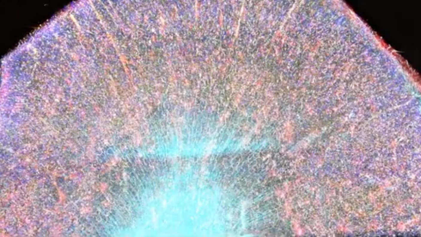
Image Credit: Image of the orbitofrontal cortex from an Alzheimer's donated brain. Chung Lab/MIT Picower Institute
Dr. Shelly Xuelai Fan is a neuroscientist-turned-science-writer. She's fascinated with research about the brain, AI, longevity, biotech, and especially their intersection. As a digital nomad, she enjoys exploring new cultures, local foods, and the great outdoors.
Related Articles

What We Actually See—and Don’t See—Shows Consciousness Is Only the Tip of the Iceberg

These Mini Brains Just Learned to Solve a Classic Engineering Problem

New Device Detects Brain Waves in Mini Brains Mimicking Early Human Development
What We Actually See—and Don’t See—Shows Consciousness Is Only the Tip of the Iceberg
These Mini Brains Just Learned to Solve a Classic Engineering Problem
New Device Detects Brain Waves in Mini Brains Mimicking Early Human Development
What we’re reading