It’s not often that a twitching, snowman-shaped blob of 3D human tissue makes someone’s day.
But when Dr. Sergiu Pasca at Stanford University witnessed the tiny movement, he knew his lab had achieved something special. You see, the blob was evolved from three lab-grown chunks of human tissue: a mini-brain, mini-spinal cord, and mini-muscle. Each individual component, churned to eerie humanoid perfection inside bubbling incubators, is already a work of scientific genius. But Pasca took the extra step, marinating the three components together inside a soup of nutrients.
The result was a bizarre, Lego-like human tissue that replicates the basic circuits behind how we decide to move. Without external prompting, when churned together like ice cream, the three ingredients physically linked up into a fully functional circuit. The 3D mini-brain, through the information highway formed by the artificial spinal cord, was able to make the lab-grown muscle twitch on demand.
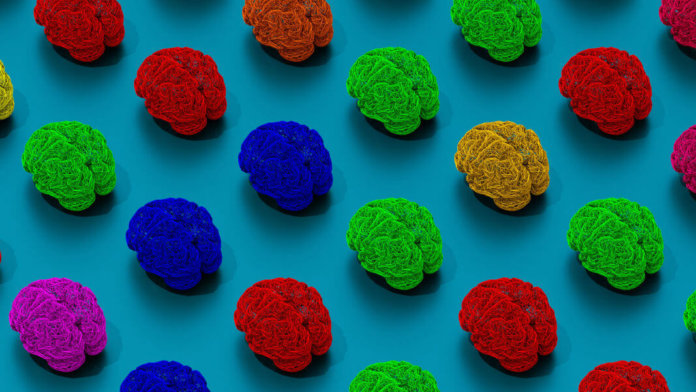
In other words, if you think isolated mini-brains—known formally as brain organoids—floating in a jar is creepy, upgrade your nightmares. The next big thing in probing the brain is assembloids—free-floating brain circuits—that now combine brain tissue with an external output.
The end goal isn’t to freak people out. Rather, it’s to recapitulate our nervous system, from input to output, inside the controlled environment of a Petri dish. An autonomous, living brain-spinal cord-muscle entity is an invaluable model for figuring out how our own brains direct the intricate muscle movements that allow us stay upright, walk, or type on a keyboard.
It’s the nexus toward more dexterous brain-machine interfaces, and a model to understand when brain-muscle connections fail—as in devastating conditions like Lou Gehrig’s disease or Parkinson’s, where people slowly lose muscle control due to the gradual death of neurons that control muscle function. Assembloids are a sort of “mini-me,” a workaround for testing potential treatments on a simple “replica” of a person rather than directly on a human.
From Organoids to Assembloids
The miniature snippet of the human nervous system has been a long time in the making.
It all started in 2014, when Dr. Madeleine Lancaster, then a post-doc at Stanford, grew a shockingly intricate 3D replica of human brain tissue inside a whirling incubator. Revolutionarily different than standard cell cultures, which grind up brain tissue to reconstruct as a flat network of cells, Lancaster’s 3D brain organoids were incredibly sophisticated in their recapitulation of the human brain during development. Subsequent studies further solidified their similarity to the developing brain of a fetus—not just in terms of neuron types, but also their connections and structure.
With the finding that these mini-brains sparked with electrical activity, bioethicists increasingly raised red flags that the blobs of human brain tissue—no larger than the size of a pea at most—could harbor the potential to develop a sense of awareness if further matured and with external input and output.
Despite these concerns, brain organoids became an instant hit. Because they’re made of human tissue—often taken from actual human patients and converted into stem-cell-like states—organoids harbor the same genetic makeup as their donors. This makes it possible to study perplexing conditions such as autism, schizophrenia, or other brain disorders in a dish. What’s more, because they’re grown in the lab, it’s possible to genetically edit the mini-brains to test potential genetic culprits in the search for a cure.
Yet mini-brains had an Achilles’ heel: not all were made the same. Rather, depending on the region of the brain that was reverse engineered, the cells had to be persuaded by different cocktails of chemical soups and maintained in isolation. It was a stark contrast to our own developing brains, where regions are connected through highways of neural networks and work in tandem.
Pasca faced the problem head-on. Betting on the brain’s self-assembling capacity, his team hypothesized that it might be possible to grow different mini-brains, each reflecting a different brain region, and have them fuse together into a synchronized band of neuron circuits to process information. Last year, his idea paid off.
In one mind-blowing study, his team grew two separate portions of the brain into blobs, one representing the cortex, the other a deeper part of the brain known to control reward and movement, called the striatum. Shockingly, when put together, the two blobs of human brain tissue fused into a functional couple, automatically establishing neural highways that resulted in one of the most sophisticated recapitulations of a human brain. Pasca crowned this tissue engineering crème-de-la-crème “assembloids,” a portmanteau between “assemble” and “organoids.”
“We have demonstrated that regionalized brain spheroids can be put together to form fused structures called brain assembloids,” said Pasca at the time.” [They] can then be used to investigate developmental processes that were previously inaccessible.”
And if that’s possible for wiring up a lab-grown brain, why wouldn’t it work for larger neural circuits?
Assembloids, Assemble
The new study is the fruition of that idea.
The team started with human skin cells, scraped off of eight healthy people, and transformed them into a stem-cell-like state, called iPSCs. These cells have long been touted as the breakthrough for personalized medical treatment, before each reflects the genetic makeup of its original host.
Using two separate cocktails, the team then generated mini-brains and mini-spinal cords using these iPSCs. The two components were placed together “in close proximity” for three days inside a lab incubator, gently floating around each other in an intricate dance. To the team’s surprise, under the microscope using tracers that glow in the dark, they saw highways of branches extending from one organoid to the other like arms in a tight embrace. When stimulated with electricity, the links fired up, suggesting that the connections weren’t just for show—they’re capable of transmitting information.
“We made the parts,” said Pasca, “but they knew how to put themselves together.”
Then came the ménage à trois. Once the mini-brain and spinal cord formed their double-decker ice cream scoop, the team overlaid them onto a layer of muscle cells—cultured separately into a human-like muscular structure. The end result was a somewhat bizarre and silly-looking snowman, made of three oddly-shaped spherical balls.
Yet against all odds, the brain-spinal cord assembly reached out to the lab-grown muscle. Using a variety of tools, including measuring muscle contraction, the team found that this utterly Frankenstein-like snowman was able to make the muscle component contract—in a way similar to how our muscles twitch when needed.
“Skeletal muscle doesn’t usually contract on its own,” said Pasca. “Seeing that first twitch in a lab dish immediately after cortical stimulation is something that’s not soon forgotten.”
When tested for longevity, the contraption lasted for up to 10 weeks without any sort of breakdown. Far from a one-shot wonder, the isolated circuit worked even better the longer each component was connected.
Pasca isn’t the first to give mini-brains an output channel. Last year, the queen of brain organoids, Lancaster, chopped up mature mini-brains into slices, which were then linked to muscle tissue through a cultured spinal cord. Assembloids are a step up, showing that it’s possible to automatically sew multiple nerve-linked structures together, such as brain and muscle, sans slicing.
The question is what happens when these assembloids become more sophisticated, edging ever closer to the inherent wiring that powers our movements. Pasca’s study targets outputs, but what about inputs? Can we wire input channels, such as retinal cells, to mini-brains that have a rudimentary visual cortex to process those examples? Learning, after all, depends on examples of our world, which are processed inside computational circuits and delivered as outputs—potentially, muscle contractions.
To be clear, few would argue that today’s mini-brains are capable of any sort of consciousness or awareness. But as mini-brains get increasingly more sophisticated, at what point can we consider them a sort of AI, capable of computation or even something that mimics thought? We don’t yet have an answer—but the debates are on.
Image Credit: christitzeimaging.com / Shutterstock.com


