The First Complete Brain Map of an Insect May Reveal Secrets for Better AI
Share
Breakthroughs don’t often happen in neuroscience, but we just had one. In a tour-de-force, an international team released the full brain connectivity map of the young fruit fly, described in a paper published last week in Science. Containing 3,016 neurons and 548,000 synapses, the map—called a connectome—is the most complex whole-brain wiring diagram to date.
“It’s a ‘wow,’” said Dr. Shinya Yamamoto at Baylor College of Medicine, who was not involved in the work.
Why care about a fruit fly? Far from uninvited guests at the dinner table, Drosophila melanogaster is a neuroscience darling. Although its brain is smaller than a poppy seed—a far cry from the 100 billion neurons that power human brains—the fly’s neural system shares similar principles to those that underlie our own brains.
This makes them excellent models to hone in on ideas of how our neural circuits wire to encode memories, make difficult decisions, or navigate social situations like flirting with a potential partner or hanging with a swarm of new friends.
To lead author Dr. Marta Zlatic at the University of Cambridge, MRC Laboratory of Molecular Biology and Janelia Research Campus,“All brains are similar—they are all networks of interconnected neurons—and all brains of all species have to perform many complex behaviors: they all need to process sensory information, learn, select actions, navigate their environments, choose food, escape from predators, etc.”
With the new connectome map, “we now have a reference brain,” she said.
A Behemoth Atlas
Connectomes are precious resources. Popularized by Sebastian Seung, the maps draw out neural connections within and across brain regions. Similar to tracing computer wires to reverse-engineer how different chips and processors fit together, the connectome is a valuable resource to crack the brain’s “neural code”—that is, the algorithms underlying its computations.
In other words, the connectome is essential to understanding the brain’s functions. It’s why similar work is underway in mice and humans, though at a much smaller scale or with far less detail.
Until now, scientists have only mapped three full-brain connectomes, all in worms—including the first animal to gain the honor, the nematode C. elegans. With just over 300 neurons, the project took over a decade, with an update released for both sexes in 2019.
Drosophila represents a far larger challenge with roughly ten times the number of neurons as C. elegans. But it’s also an ideal next candidate. For one, scientists have already sequenced its entire genome, making it possible to match genetic information to the fly’s neural wiring. This could especially come in handy for, say, deciphering how genes contributing to Alzheimer’s disease alters neural circuits. For another, fruit fly larvae have transparent bodies, making them far easier to image under a microscope.
Not all brain-wiring maps are created equal. Here, the team went for the highest resolution: mapping the whole brain at the synapse level. Synapses are junctions between neurons where they connect: picture two mushroom-shaped structures hovering near each other with a gap. Although neurons are often touted as the basic component of computing, synapses are where the magic happens—their connectivity helps functionally wire up neural circuits.
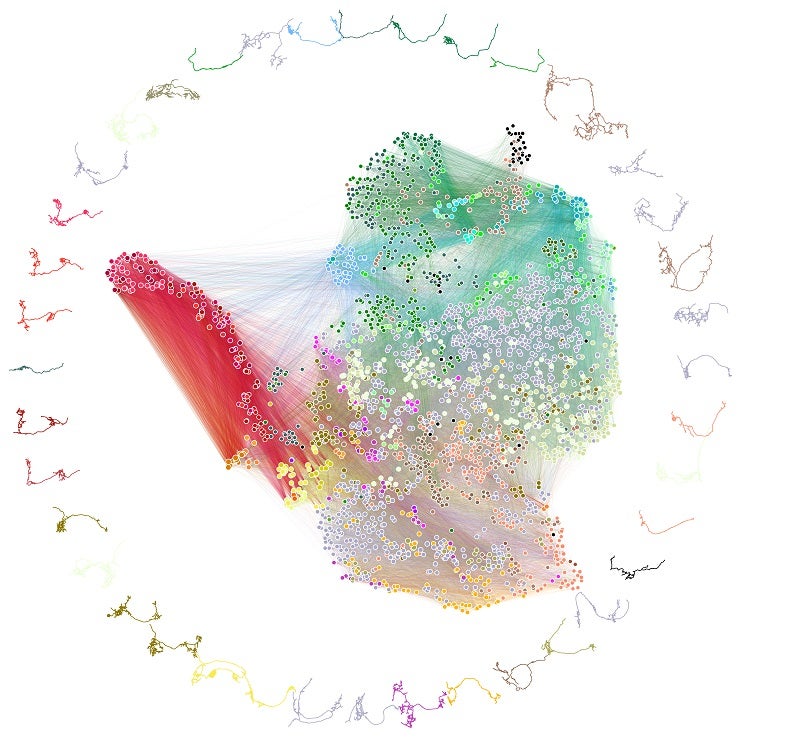
Neuron connectivity in the brain. Each dot represents a neuron, and those with more similar connectivity are closer. The lines show how neurons connect. Image Credit: Benjamin Pedigo
Slice and Dice and…Robots?
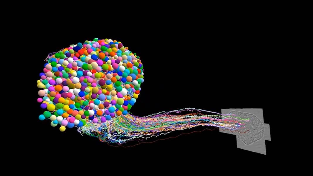
To map out synapses, the team turned to the big guns of microscopy: the electron microscope. Compared to microscopes in high school biology, this hardware can capture images at the nanoscale—roughly a tenth the width of a human hair.
The whole process sounds a bit like a wild dinner recipe. The team first soaked a single six-hour-old larvae brain inside a solution packed with heavy metals, which marinated into the neurons’ membranes and proteins inside synapses. The brains were then painstakingly sliced into ultra-thin sections with a diamond blade—imagine a deli-meat slicer—and put under a microscope.
The resulting images—all 21 million of them—were stitched together using software. The whole process took over a year and a half, with many hours spent manually checking the reconstructed neurons and synapses.
The final brain map didn’t just contain the location of neurons and their synapses—it also highlighted wiring quirks that could support highly efficient neural computations.
Winding Roads
The beauty of the new map is that it provides bird’s-eye information on brain connectivity, supercharged with the power of zoom-and-enhance.
“The most challenging aspect of this work was understanding and interpreting what we saw,” said Zlatic.
In one analysis, the team found that neurons can be grouped into 93 different types based on their connectivity, even if they share the same physical structure. It’s a drastic departure from the most common way of categorizing neurons. Rather than clustering them based on appearance or function, it may be more useful to focus on their connectivity “social network” instead.
Be Part of the Future
Sign up to receive top stories about groundbreaking technologies and visionary thinkers from SingularityHub.

Digging down to synapses, the team ran into another surprise. Let me explain: neurons have two main branches. One is the larger input cable—the axon—and the other is a tree-shaped output—the dendrite. Neurons usually “wire up” when synapses connect those two cables.
More recent studies, however, show that synapses on axons can connect with other synapses on axons; the same goes for dendrites. Analyzing the reconstructed brain, the team found evidence of these non-traditional connections.
“Now we need to reconsider them: we probably need to think about creating a new computational model of the nervous system,” said Dr. Chung-Chuang Lo at the National Tsing Hua University in Taiwan.
On a broader scale, the map showed that neurons are eager to chat with others a half-world away. Almost 93 percent of neurons connected with a partner neuron in the other brain hemisphere, suggesting that long-range connections are incredibly common. Even more surprising was a peculiar population that didn’t reach out: dubbed Kenyon cells, these neurons mostly populate the fly’s learning and memory center. Why this happens is still unclear, but it illustrates the brain map’s ability to generate new insights and hypotheses.
Although the neurons and synapses are wired in a nicely compact “nested” multilayered structure, the connectome showed that some developed connections that jumped through layers—a shortcut that hooks up otherwise separate circuits.
Even more fascinating was how much the brain “talks” to itself. Nearly 41 percent of neurons received recurrent input—that is, feedback from other parts of the brain. Each region had its own feedback program. For example, information generally flows from sensory areas of the brain to motor regions, although the reverse also happens and creates a feedback loop.
But perhaps the most socially adept neurons are those that pump out dopamine. Well known for encoding reward and driving learning, these neurons also had some of the most complex recurrent wirings compared to other types.
From shortcuts to recurrent wirings, these biological hardware structures could increase the brain’s computational capacity and compensate for the limited number of neurons and their biological restraints.
“None of us expected this at all,” said study author Dr. Michael Winding.
From Fly to AI
The study isn’t the first to map the Drosophila brain. Previously, a team led by Dr. Davi Bock at the Janella Research Campus targeted a small nub of the adult fruit fly brain responsible for learning and remembering smells with synapse-level detail. Zlatic’s team has also tracked a sensory circuit in the fruit fly larvae for making decisions by mapping only 138 neurons.
The full-brain connectome is a game-changer. For one, scientists now have a sophisticated reference brain to test out theories for neural computation. For another, the connectome map and its inferred computation resembles state-of-the-art machine learning.
“That’s really quite nice because we know that recurrent neural networks are pretty powerful in artificial intelligence,” said Zlatic. “By comparing this biological system, we can potentially also inspire better artificial networks.”
Image Credit: Michael Winding
Related Articles

Scientists Find a Surprising New Way Stress Cascades From Brain to Body

How the Bilingual Brain Switches Languages With Ease

Woman With Alzheimer’s Shows Striking Improvement After Taking Magic Mushrooms
Scientists Find a Surprising New Way Stress Cascades From Brain to Body
How the Bilingual Brain Switches Languages With Ease
Woman With Alzheimer’s Shows Striking Improvement After Taking Magic Mushrooms
What we’re reading